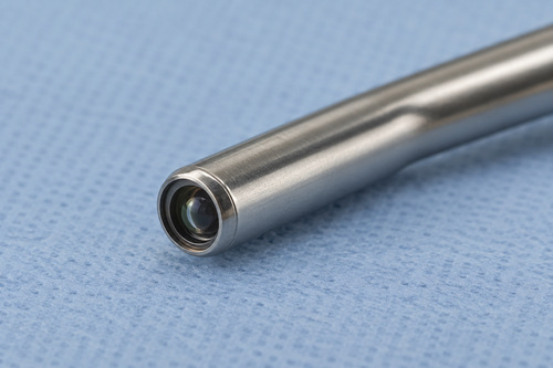

A clear endoscopic image depends on more than the camera sensor. Light has to reach the tissue, return through the viewing optics, and pass through surfaces that remain usable during the procedure. Windows, lenses, fibers, coatings, and their joints all influence that path.
For reusable equipment, those materials also have to remain compatible with the instrument’s specified cleaning and reprocessing conditions. A component can transmit light well when new and still be unsuitable for the way the assembled device will be handled. Optical performance and service conditions therefore belong in the same material-selection discussion.
This is especially relevant to small instruments, where a window may perform several tasks at once. It can transmit the imaging signal, protect internal optics, and form part of the barrier between the device interior and its surroundings.
Different optical parts have different jobs
A lens forms or relays an image. An optical fiber carries illumination or another optical signal. A window generally passes light while separating regions of the assembly. Although all may be transparent, they are specified differently.
The window’s clear aperture must suit the useful viewing field. Its thickness, surface shape, and relationship to the surrounding optics must also be considered. A protective disc selected only for outer diameter can fit the housing while changing the path of the imaging rays.
Material choices may include optical glasses, fused silica, and crystalline materials. Sapphire windows are one established option where their optical and mechanical properties suit the instrument. In a published laryngeal imaging design, a sapphire window formed part of a sealed assembly protecting an internal optical pattern. [1]
The useful lesson is the component’s role in the device. It is an optical part and an assembly interface, so both functions need to appear in the drawing.
Match transmission to the imaging method
Ordinary white-light viewing is only one endoscopic technique. Specialized instruments can use selected spectral bands, fluorescence, or polarization-sensitive measurements. The material must transmit the relevant illumination and return signals.
A window that appears clear to a person can behave differently outside the visible spectrum. Thickness and material grade can affect absorption. Coatings can further change transmission, particularly away from their design wavelength or angle.
If illumination and detection use different bands, both need to be specified. Otherwise, a component may transmit the excitation light adequately while weakening the signal the instrument is meant to collect.
This also affects replacement parts. Matching the old component’s outline is insufficient if its material grade, coating, or optical orientation is unknown. The replacement specification should capture the behavior that matters to the original optical design.
Polarization creates a specific material requirement
Polarization-sensitive methods analyze more than the brightness of reflected light. They depend on preserving or deliberately controlling its polarization state along the optical path.
Sapphire is birefringent: its response depends on crystallographic direction and polarization. A window can consequently introduce a change that the instrument must account for. Research on polarized endoscopy has measured these effects and examined alternative window arrangements. [2]
One polarization-enhanced laparoscope used a fused-silica window to avoid effects associated with the conventional sapphire component. That choice followed the requirements of a particular measurement method. It does not mean that sapphire is unsuitable for ordinary endoscopic viewing. [3]
For the buyer, the key question is whether the instrument is sensitive to polarization and over what range of viewing angles. If orientation matters, the drawing should identify it rather than leaving the supplier to interpret a general requirement for sapphire.
Surface condition affects the usable view
Scratches, contamination, and poorly controlled coatings can scatter or redirect light. These effects may reduce contrast or introduce unwanted reflections even when a window remains visibly transparent.
Surface quality and flatness describe different characteristics. A cosmetic defect requirement addresses one type of surface condition; it does not establish the component’s transmitted optical performance. The appropriate specification depends on the imaging path and the useful aperture.
Edges and mounting lands need their own attention. They can be outside the image-forming region yet remain essential to seating or sealing the part. A chip at a sealing interface may matter for reasons quite different from a scratch in the clear aperture.
Where a stepped geometry helps the mechanical design, a sapphire step window is a distinct component form to consider. A step needs a drawing defining its dimensions and mating surfaces; it should not be assumed to provide a particular seal without an assembly design.
The joint must survive the intended processing
A durable substrate does not establish the durability of everything attached to it. Adhesives, coatings, seals, and surrounding metals may respond differently to temperature and chemicals.
The FDA describes reusable-device reprocessing as device-specific. Instructions and their validation apply to the finished equipment and its intended sequence of cleaning, disinfection, or sterilization. [4]
For development, those conditions provide useful component requirements. The engineering team can identify the exposures the window assembly will experience and evaluate whether its optical and mechanical behavior remains acceptable afterward.
This is not a reason to choose the hardest material by default. It is a reason to examine the whole interface. A substrate that performs well on its own can still be part of an assembly with a vulnerable bond or coating.
Keep condensation separate from material transparency
A window can have suitable bulk transmission and still provide a poor view if moisture condenses on an exposed surface. That is a service condition of the assembled instrument, rather than a change in the basic identity of the substrate.
Published evaluation of a polarization-enhanced laparoscopy prototype reported imaging failures that included front-window condensation. The example shows why optical assessment needs to include the working environment, even after the material has been selected for an appropriate polarization response. [5]
During development, the team can identify which side of the window faces the examination environment, whether the opposite side encloses air, and what temperature changes occur during use. Those details help guide the assembly review without assuming that a different transparent material will resolve the issue by itself.
If a coating or other surface treatment is proposed, its optical effect and compatibility with the instrument’s processing requirements also need evaluation. A successful material choice and a controlled operating interface have to work together.
Discuss the component with a material supplier early
Stanford Advanced Materials (SAM) supplies optical crystal materials for medical equipment, including sapphire and other window materials. A useful inquiry describes the wavelength band, dimensions, clear aperture, optical surfaces, crystal orientation where relevant, and intended mounting method.
If the part will be joined or coated elsewhere, identify those later steps. The supplied blank may need particular dimensions or surface conditions to allow the downstream process. Discovering that requirement after delivery can create unnecessary rework.
The expected quantity and development stage are also useful. A prototype order may leave some dimensions open for discussion, while a repeat component may need to follow an established revision. Separating fixed requirements from adjustable ones helps the supplier review practical options.
Keep inspection connected to the complete optical path
Incoming checks can confirm dimensions, surface condition, and other agreed component requirements. The finished instrument then needs the optical measurements relevant to its function.
For example, a development team investigating poor image quality can compare the unmounted component with the assembled optical path. That can help distinguish a material or surface problem from a mounting, illumination, or alignment problem. A photograph showing a clear window cannot make that distinction.
For sourcing assistance, SAM can review material and component requests against the supplied drawing and specification. The strongest request explains what the window has to transmit, protect, and fit. Those details connect material selection to the usable view the clinician ultimately needs.
References
[1] Biomedical Optics Express. Laser-Calibrated System for Transnasal Fiberoptic Laryngeal High-Speed Videoendoscopy
[2] Biomedical Optics Express. Polarized multispectral imaging in a rigid endoscope based on elastic light scattering spectroscopy
[3] Biomedical Optics Express. Polarization enhanced laparoscope for improved visualization of tissue structural changes associated with peritoneal cancer metastasis
[4] U.S. Food and Drug Administration. Reprocessing Medical Devices in Health Care Settings Validation Methods and Labeling
[5] Biomedical Optics Express. Evaluation of a polarization-enhanced laparoscopy prototype for improved intra-operative visualization of peritoneal metastases